Have you ever wondered how the brain works? At the Brain Foundation we talk a lot about brain diseases, disorders, and injuries, often sharing research updates and medical news. But if you don’t understand the basic structure and function of the brain, we know that some of these topics can seem quite complicated.
In this article, we explain some of the basics about how the brain works. This was initially published in a three part series in our biannual Brainwaves newsletter.
Click the headings below to jump to a specific section.
Introduction
The human brain is the centre of the body’s nervous system and the locus of your cognition. It is responsible for everything that you do, feel, and perceive. So when you are diagnosed with a brain disease, disorder, or injury, it can be quite distressing news.
We talk about these conditions quite a lot at the Brain Foundation. But have you ever wondered how the brain works in the first place? Maybe you have questions like…
- How do I learn & process information?
- What is the difference between white matter & grey matter?
- How is the physical & biological structure of the brain connected to different brain diseases, disorders, and injuries?
Learning these brain basics can help you understand more about neurological conditions, and help you decode some of the scientific jargon you might see in medical articles. We hope this brief introduction will help make neuroscience seem a little bit less intimidating.
What is a neuron?
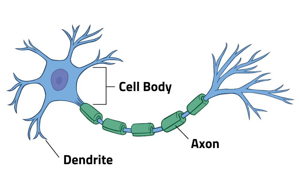
The central nervous system (CNS) is made up of two types of cells – neurons (or nerve cells) and glia. Neurons are the key players in the brain. They are the information messengers that allow us to think, learn, move, and feel; while glial cells primarily support and protect neurons.
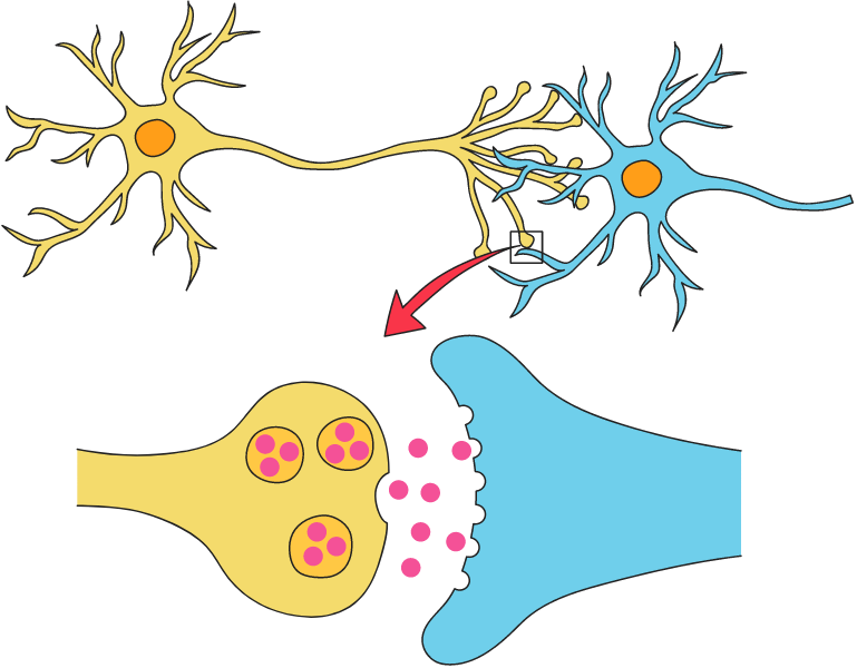
There are many different types of neurons, but they all consist of three key parts.
These cell functions (input, output, and processing) are controlled by electrical and chemical signals. If a neuron is receiving enough information through its dendrites, it will respond by sending an impulse down the axon.
At the end of the axon, the cell will emit neurotransmitters or electrical signals through the synapse to be received by another neuron. These messages are being passed around constantly, allowing you to experience, understand, and react to the world.
White matter & grey matter
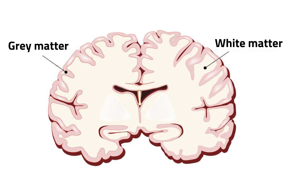
Grey and white matter are the two regions of the central nervous system. They are made up of different cells, and serve different purposes.
Grey matter is the part of your brain that does most of the actual processing, thinking, and interpreting information. It is mostly made up of neuronal cell bodies with shorter axons.
White matter, on the other hand, is the connective tissue of your nervous system. It is composed mostly of myelinated axons, which transport messages between different parts of your brain and body. The neurons in white matter still have cell bodies, but they have much longer axons. The longest axons are in the dorsal root ganglion, and they stretch from your toes to your brainstem (up to two metres in a tall person)!
These regions are relevant when talking about specific brain diseases. Neurodegenerative diseases such as Alzheimer’s disease and Parkinson’s disease are caused by neuron loss in the grey matter. White matter might also be altered, but the main disease markers (such as amyloid plaques) are located in the grey matter.
White matter diseases affect nerve signal transmission, which can cause serious issues. Multiple sclerosis is one example of white matter disease. It occurs when the myelin that coats your axons is destroyed, leading to motor or sensory disruption.
Main parts of the brain
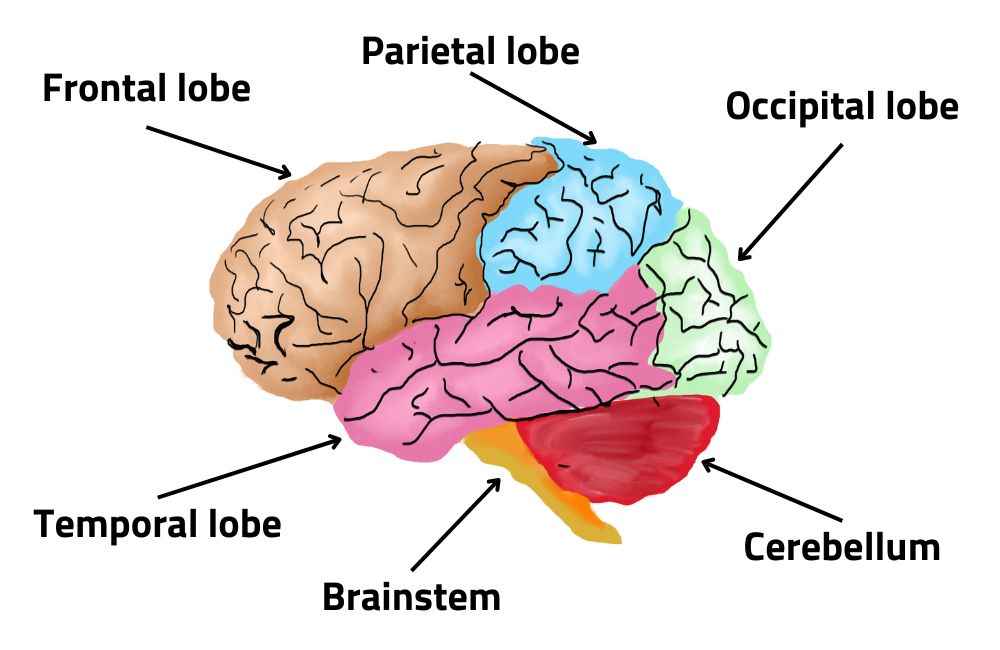
There are three main parts of the brain: the cerebrum, the cerebellum, and the brainstem.
Learning about the function of these regions in the brain can help you understand how the brain works together as a whole, and will hopefully demystify some of the scientific jargon you might see in medical articles.
Cerebrum
The cerebrum is the largest part of the brain and it performs higher levels of thinking and action. This includes speech, judgement, emotions, learning, reasoning, and interpreting touch, vision & hearing. It is made up of four lobes which each perform a different job.
- The frontal lobe: This is at the front and top of the brain. It is responsible for tasks that are essential in higher level thinking and behaviour, such as planning, judgement, decision making, impulse control, and attention.
- The parietal lobe: The parietal lobe is behind the frontal lobe. It takes in sensory information (including pain and temperature) and spatial information, which help you understand your position in your environment.
- The temporal lobe: This is at the lower front of the brain. It is important for visual memory, emotion, and language.
- The occipital lobe: This lobe is at the back of the brain, and it processes visual input from the eyes.
Cerebellum
The cerebellum might look small, but it actually contains as many as 80% of all neurons in the brain! It is located under the cerebrum, and is responsible for coordinating muscle movements, maintaining posture, and balance.
These are complex functions involving special sensors that detect shifts in balance and movement. These sensors (alongside other parts of the brain) help you do everything from standing upright to learning complex motor skills like riding a bike.
New studies are also exploring the cerebellum’s role in thought, emotions, and social behaviour. This might reveal some involvement in psychiatric and neurological disorders.
Brainstem
The brainstem is at the bottom of the brain and connects the cerebrum & cerebellum to the spinal cord. It controls many vital automatic functions, such as breathing, circulation, and sleeping. It is composed of:
- The midbrain: This is a complex part of the brainstem, and it facilitates many functions. Some of these include hearing, movement, and calculating responses to changes in your environment.
- The pons: The pons is the origin of four cranial nerves. It is responsible for many facial movements and functions, such as chewing, blinking, focusing your eyes, tear production, and facial expressions.
- The medulla: This is where the brain meets the spinal cord. The medulla is critical for survival, as it regulates your heart rhythm and breathing. It also controls reflex activities such as coughing or sneezing.
Involvement in brain disorders
Now that we’ve explained how these parts of the brain work, we can start to think about how brain diseases, disorders, and injuries can affect them.
Some brain diseases can occur in any part of the brain, such as stroke. Other conditions only affect a specific region, which is reflected in the symptoms. And in other cases, it might be more complex. For example, dystonia is not a single disease but a syndrome – a set of symptoms that cannot be attributed to a single cause but share common elements.
As a general guideline, these are some of the symptoms you might expect to see depending on what brain region is affected. Keep in mind that this is a very simple overview – brain diseases can affect people differently or develop in unexpected ways.
| Region | Possible symptoms | Related conditions |
| Cerebrum | Huge variety of symptoms, particularly depending on what lobe is affected. Could include motor impairments, problem solving and judgement issues, behavioural changes, and difficulty with speech. | Cerebral palsy, frontotemporal dementia, epilepsy. |
| Cerebellum | Lack of muscle control and coordination, walking & mobility issues, slurred speech, abnormal eye movements. | Friedreich’s ataxia & other types of ataxia, Duchenne muscular dystrophy. |
| Brainstem | Coma, difficulty breathing, difficulty swallowing, sleep problems, apathy, trouble regulating blood pressure. | Parkinson’s disease, Locked-in syndrome & other disorders of consciousness, Basilar migraine. |
Deeper structures in the brain
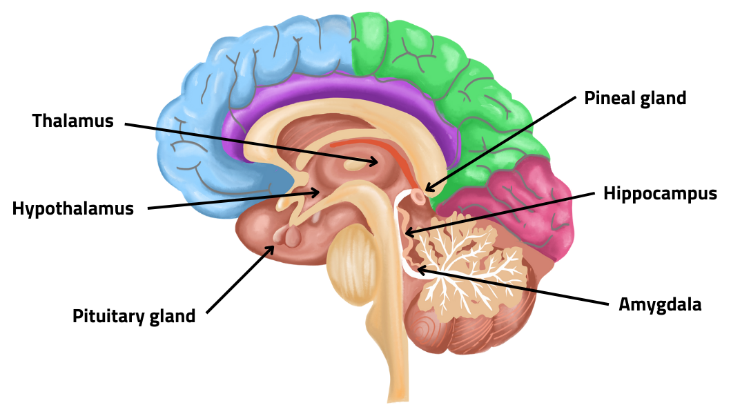
The cerebrum, cerebellum and brainstem are the largest parts of the brain. These are the sections that you could see from the outside if you were to look at a model or preserved brain. However, there are other structures that are not visible from the outside.
These structures may be small compared to the wrinkly outer layers of the brain, but they are essential for human survival. They work together to regulate vital bodily functions, memory, and emotional responses. Dysfunction in any of these structures can lead to a variety of neurological disorders, including memory loss, sleep disorders, and hormonal imbalances.
Hypothalamus
The hypothalamus is a small structure located at the base of the brain. It controls the involuntary physiologic processes (the autonomic system). It plays a crucial role in regulating behaviours such as body temperature, hunger, thirst, sleep, and sexual response. It also controls the release of hormones from the pituitary gland.
Thalamus
The thalamus is located in the centre of the brain and relays sensory information between your body and the cortex. It receives information from various sensory organs such as your eyes, ears, and skin. It plays a role in regulating consciousness, attention, sensing pain, and memory.
Pituitary gland
The pituitary gland is a pea-sized gland in the base of the brain. It is also known as the ‘master gland’ because it controls other endocrine glands in the body, so it is responsible for regulating most of our hormones. It secretes hormones that control sexual development, growth, and metabolism.
Pineal gland
The pineal gland is another small gland deep in the brain. It is crucial for regulating our internal clock because it secretes melatonin. Melatonin is produced when it gets dark, and it is the hormone that controls sleep and wake cycles.
Hippocampus
The hippocampus is a curved structure on the underside of the temporal lobe. It is responsible for forming and storing long-term memories. It also supports learning, spatial perception, and navigation. Research shows that damage to the hippocampus may play a role in Alzheimer’s disease.
Amygdala
There are two amygdalae in the brain, one under each hemisphere. They regulate memory and emotions. The amygdala is also associated with the brain’s reward system, stress, and the ‘fight or flight’ response.
Neurotransmitters
Neurotransmitters are chemical messengers in the brain. They allow neurons to communicate with each other or with other cells in the body. Neurotransmitters are released out of the axon of a neuron into a small gap between cells, called a synapse, then bind to a receptor on the other cell.
There are at least 100 different neurotransmitters, each with their own specific role. There are three main types of neurotransmitters. Excitatory neurotransmitters create electrical signals that encourage the target cell to take action. Inhibitory neurotransmitters stop cells from being activated. Modulatory neurotransmitters can influence multiple target cells at once, and also regulate the effect of other neurotransmitters.
Examples of common neurotransmitters
Acetylcholine: Regulates muscle contractions and is involved in cognitive functions such as memory and learning. Low levels of acetylcholine are linked to memory loss and Alzheimer’s disease.
Glutamate: The most common & powerful excitatory neurotransmitter. It has many functions and plays a key role in learning, memory, and mood regulation. Glutamate imbalances are linked to many disorders such as epilepsy, neurodegenerative disease & mood disorders.
Serotonin: An inhibitory neurotransmitter that helps regulate mood, behaviour, sleep, and memory. Low serotonin levels are linked to depression, anxiety, and sleep disorders.
Norepinephrine: An excitatory neurotransmitter that is crucial to the body’s stress response. It increases heart rate & blood pressure in response to fear or stress. It affects your alertness, arousal, decision-making, attention and focus. Abnormal levels of norepinephrine are linked to psychiatric disorders and Parkinson’s disease.
Further information & resources
We hope this article helped you learn a bit more about how the brain works. If you’d like to learn more about the brain or any specific disorders, you can visit the following pages:
- Brain Disorders A-Z – our directory of over 70 brain diseases, disorders and injuries
- Healthy Brain – learn about brain health & tips to maintain brain health
- What is brain disease? – this article explains what brain disease is, including some of the common types of brain diseases, disorders and injuries.
- Newsletter – subscribe to our newsletter for regular updates


 The Brain Foundation is the largest, independent funder of brain and spinal injury research in Australia. We believe research is the pathway to recovery.