
Combined imaging in the diagnosis of early Corticobasal Syndrome
Associate Professor John O’Sullivan
Royal Brisbane & Women’s Hospital
Co-Investigators: Dr Andre Troiano, Ms Amy Jones, Associate Professor Stephen Rose and Dr David MacFarlane
Corticobasal degeneration (CBD) is an uncommon but devastating neurodegenerative condition. Patients develop progressive problems with movement and cognitive function, typically affecting one side of the body first. Many patients who appear to have the typical clinical features of CBD are found at post mortem to have pathological changes of different diseases such as Parkinson’s disease(PD), frontotemporal dementia(FTD) and progressive supranuclear palsy(PSP). Conversely patients with CBD at post mortem may present with clinical features more suggestive of these other diseases. Because of this discrepancy, the clinical features are referred to as corticobasal syndrome (CBS) and CBD is reserved for those who have pathological confirmation. Providing accurate diagnosis and prognosis for patients with early symptoms of CBS is very difficult.
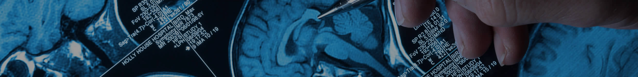
Advanced brain imaging techniques utilizing magnetic resonance imaging (MRI) and positron emission tomography (PET) reveal some differences between these conditions in established cases, but it is unclear if such techniques will help in the early differentiation of these conditions.
In this study, patients with early and inconclusive early features of CBS will be examined in detail and undergo MRI and PET imaging. After 3 years, the patients will be examined again, and with progression of the disease a more definitive clinical diagnosis will be possible. We will then determine whether changes on the original scan were helpful in determining the final diagnosis. We also hope to obtain a final pathological diagnosis in patients after death.
With research suggesting different treatments to help slow the progression of distinct neurodegenerative diseases, it will become critical to more accurately differentiate neurodegenerative diseases as early as possible to appropriately target therapy and to provide a more accurate prognosis. This results of this project will determine whether MRI and PET imaging will be useful in the early diagnosis of these conditions.